WBF What is
brain fog?
Support WBF Take quiz
Medically reviewed by Dr. Alexandru-Theodor Amarfei, M.D.

EEG and Brain Fog: What Brain Waves Reveal

Key Takeaways

  • Group-level EEG differences: small 2024 studies report average changes in post-COVID and ME/CFS groups, not a test for individuals
  • Three reported changes: less posterior alpha (8-13 Hz), more theta (4-7 Hz) and slower P300 responses, all of which also appear with fatigue and poor sleep
  • Not clinically standard yet: Research shows promise but routine EEG testing for brain fog isn't established
  • Consumer devices can't detect this: Muse, DREEM, etc. lack the resolution and electrode coverage
  • EEG results may vary by condition: Long COVID, fibromyalgia and Alzheimer's look different

EEG research today

For decades, people with brain fog have often heard that their routine tests look normal. Recent EEG research suggests some groups of these people may show measurable brain wave differences on average. But it's still new and isn't a standard clinical answer.

What is EEG?

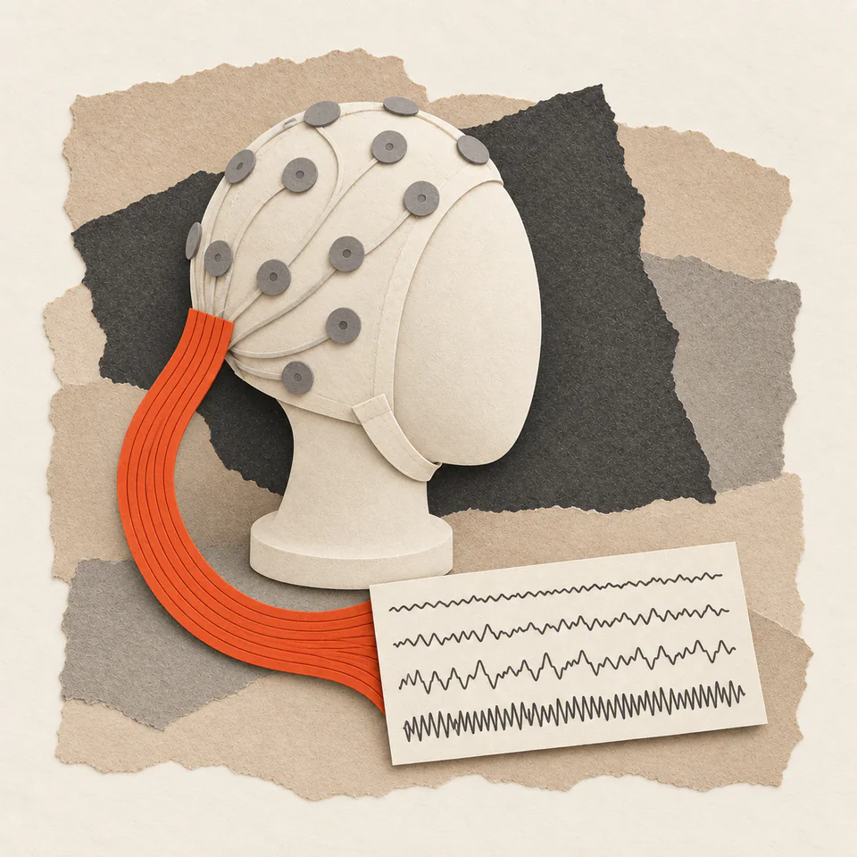
Electroencephalography (EEG) measures the electrical activity of your brain using electrodes placed on the scalp. When neurons communicate, they create tiny electrical signals. These signals add up to rhythms called "brain waves," which EEG records live.

Unlike MRI, which shows brain structure, EEG shows brain function: what your neurons are doing moment to moment. That makes it useful for research on how brain activity shifts in brain fog.

Standard vs. quantitative EEG

  • Standard EEG: Used clinically for epilepsy, sleep disorders, coma evaluation. Neurologists read it by eye for abnormal activity. Brain fog doesn't typically show obvious abnormalities on standard EEG.
  • Quantitative EEG (qEEG): A computer compares your brain waves with normal reference data. This is what research studies use to detect subtle brain fog changes. Sometimes called "brain mapping."

Clinical EEG uses 19-64 electrodes in a standard layout (the 10-20 international system). Research setups may use 128-256 for finer detail.

Brain wave basics

Brain wave types differ by frequency (cycles per second, measured in Hertz/Hz). Different frequencies dominate during different mental states:

Wave Type Frequency Associated State
Delta 0.5-4 Hz Deep sleep, healing, unconscious processes
Theta 4-7 Hz Drowsiness, light sleep, meditation, memory consolidation
Alpha 8-13 Hz Relaxed and awake, ready to engage
Beta 13-30 Hz Active thinking, concentration, problem-solving
Gamma 30-100 Hz Higher cognitive functions, binding information

A healthy brain shifts between these states fluidly. In brain fog, group averages suggest this flexibility may be reduced, with less power in some frequencies and more in others.

What the 2024 studies found

Recent studies have started converging on similar findings in Long COVID, ME/CFS, and related cognitive syndromes.

Babiloni et al. (2024): Long COVID EEG

Journal: Clinical Neurophysiology | Sample: post-COVID adults with brain fog, about one year after hospital admission, versus matched controls

Key Finding: The post-COVID group had less resting alpha activity at the back of the brain than the comparison group, especially people with two or more fatigue symptoms. The alpha change accompanied the brain fog and fatigue people reported. More than 90% had no cognitive or psychiatric disorder.

PMID 38492271. DOI: 10.1016/j.clinph.2024.02.034

Gangemi, Suriano and Fabio (2024): P300 study over time

Journal: J Integrative Neuroscience | n= 40 (brain fog with and without smell loss, plus controls), EEG at 1 and 8 months after COVID-19 recovery

Key Finding: People with brain fog after COVID-19 showed different P300 and beta-band measures, and the differences were still there eight months after recovery from the infection.

PMID 38812399. DOI: 10.31083/j.jin2305105

Silva-Passadouro et al. (2024): Systematic review of qEEG in fibromyalgia, ME/CFS and Long COVID

Journal: Clinical Neurophysiology | Scope: 17 resting-state qEEG studies compared with healthy controls

Key Finding: Fibromyalgia studies tended to show less low-frequency (delta, theta, alpha) and more beta activity. ME/CFS looked different, and the few Long COVID studies gave mixed results. The reviewers treated these as research leads, not a way to diagnose.

PMID 38772083. DOI: 10.1016/j.clinph.2024.04.019
Less alpha activity at rest

Alpha waves (8-13 Hz), especially at the back of the brain (visual cortex, parietal regions), show the brain is relaxed but ready. At this frequency, it can engage quickly when needed.

In Long COVID and ME/CFS brain fog, posterior alpha power is reduced. This may explain several characteristic symptoms:

  • Trouble starting tasks: Without enough resting alpha activity, the brain struggles to shift into focus
  • Mental fatigue: The brain may work harder to function normally and tire faster
  • Reduced mental clarity: Alpha waves appear during calm, focused states, so less alpha means more distraction

Overlap with Alzheimer's

Reduced posterior alpha is also common in Alzheimer's disease and mild cognitive impairment (MCI). So a single EEG needs careful reading. The overlap isn't evidence that post-viral brain fog leads to dementia. EEG can't tell these apart. Clinical assessment and cognitive testing over time can.

P300 delay: slower information processing

The P300 is an "event-related potential," a brain response that comes about 300 milliseconds after you notice something significant (hence the name). It's measured using an "oddball" task: you hear a series of beeps (boop-boop-boop) and occasionally a different tone (BEEP). Your brain generates a P300 wave when it recognizes "that was different."

P300 latency measures how long this recognition takes. In brain fog conditions:

  • Normal P300: ~300ms latency
  • Reported in study groups: later or smaller P300 responses on average. The numbers vary by lab, task and age
  • What it means: on average these groups processed the stimulus more slowly. A single P300 result is not a brain-fog test

The Gangemi, Suriano and Fabio study found that people who rated their brain fog as worse had longer P300 latencies. In the brain fog group, the P300 differences were still there eight months after recovery from COVID-19.

What P300 can show

P300 depends less on self-report than a questionnaire, but it still varies with age, alertness, attention and medication, and there is no agreed brain-fog cut-off. A delayed P300 says processing was slower in that session. It does not say why.

More theta: drowsy while awake

Theta waves (4-7 Hz) normally dominate during drowsiness, light sleep, and the transition between waking and sleeping. Some theta while you're awake is normal. It's linked to forming memories and creative thinking.

But excessive theta during wakefulness suggests the brain is operating in a drowsy state despite being "awake." This shows up in:

  • Sleep deprivation (your brain trying to sleep while you force it awake)
  • Attention disorders (ADHD shows increased theta/beta ratio)
  • Brain fog conditions (extra theta in tasks needing alertness)

Researchers see the extra theta in brain fog as the feeling of being "half-asleep," unable to fully engage with tasks that need alertness. Theta also rises with drowsiness and sleep loss.

EEG changes by condition

"Brain fog" is a symptom, not a diagnosis. Different conditions may produce different EEG changes:

Condition Main EEG changes
Long COVID / ME/CFS Reduced posterior alpha, increased theta, delayed P300 (group averages in small studies)
Fibromyalgia Decreased low-frequency power, increased beta (different from ME/CFS)
Alzheimer's/MCI Progressive alpha slowing and theta increase over time
ADHD Elevated theta/beta ratio (excess slow waves relative to fast)
Depression Frontal alpha asymmetry (greater right-sided activation)

These differences suggest that brain fog from different causes may need different treatment, even when the symptoms feel alike.

Clinical use today

Despite promising research, routine EEG testing for brain fog isn't clinically standard yet. Here are four reasons:

1. No standard clinical method

Research studies use specific methods (eyes-closed rest, P300 oddball tasks) that aren't part of standard clinical EEG. Most neurologists ordering an EEG are looking for seizures or encephalopathy, not subtle alpha/theta changes.

2. Conclusions vary

qEEG compares readings with normal reference databases. Different databases, different electrode setups, and different analysis methods can yield different conclusions. There's no gold standard yet.

3. Insurance coverage

Standard EEG may be covered; qEEG "brain mapping" often isn't. Out-of-pocket costs for full qEEG assessment range from $500-2,000.

4. Rarely changes treatment

Even if a qEEG report shows results like those in the studies above, it doesn't confirm brain fog, and there's no FDA-approved treatment that targets these EEG changes. The report may feel validating, but it rarely changes your care.

When standard EEG makes sense

If you have brain fog with sudden onset, seizure-like episodes, fainting, sudden personality changes or neurological symptoms in one area, your doctor may order a standard EEG. It checks for epilepsy, encephalitis (brain inflammation) or other serious conditions. That's useful even if it doesn't detect "brain fog."

Consumer EEG Devices

Consumer EEG headbands (Muse, Neurosky, DREEM, etc.) have become popular for meditation tracking and sleep monitoring. Can they detect brain fog changes?

No, they can't

Consumer devices can't replicate the clinical findings from research studies for several reasons:

  • Electrode coverage: Consumer devices use 2-7 electrodes; clinical EEG uses 19-64. Researchers measure alpha at the back of the head, which consumer devices don't cover.
  • Recording quality: Dry electrodes, movement and limited amplification make the readings much noisier than clinical wet-electrode setups.
  • No P300 capability: Consumer devices don't have the stimulus presentation and timing precision needed for event-related potentials.
  • No reference comparison: They show your data without comparing it to validated databases of healthy and brain fog readings.

Consumer EEG devices may be useful for meditation practice, sleep stage tracking or general biofeedback. They aren't suitable for diagnosing or ruling out brain fog or neurological conditions, or for judging treatment effects.

What this means for you now

1. Validation

Someone may have told you brain fog is "just anxiety" or "all in your head." Small studies have reported group-level EEG differences in some post-viral groups. Your experience does not need an EEG to be valid, and a normal EEG does not make it less real.

2. Months-long changes

The Gangemi 2024 follow-up found that P300 and beta changes were still present eight months after COVID-19, so recovery on EEG is neither quick nor guaranteed. Symptom recovery is monitored clinically, not by EEG.

3. Hold off on qEEG

Unless you have red flags needing neurological evaluation, paying out-of-pocket for qEEG brain mapping is unlikely to change your care. The money may be better spent on treatments with solid evidence (addressing sleep, nutrition, inflammation).

4. Still research-only

EEG remains a research tool in brain fog. No EEG-based diagnostic or treatment protocol exists for brain fog today. We will update this section if that changes.

FAQ

Should I ask my doctor for an EEG?

If you have brain fog without red flags (seizures, sudden onset, loss of consciousness), a routine EEG is unlikely to help. It's designed to detect epilepsy and encephalopathy, not subtle brain fog changes. But if your doctor suspects other conditions, an EEG may be appropriate to check for them.

What about neurofeedback?

Neurofeedback has not been tested in brain fog trials. It uses real-time EEG feedback to train particular rhythms, and the studies that exist are small, mostly in other conditions such as ADHD, and rarely replicated. There is no validated brain-fog protocol to look for. If you are considering it, treat it as an unproven, self-funded intervention and put the money towards a diagnostic work-up first if you have not had one.

Can Muse or other consumer devices detect my brain fog?

No. Consumer devices lack the electrode coverage, recording quality and analysis tools to detect the changes seen in research studies.

Does reduced alpha mean I have early dementia?

No. Similar EEG changes can come from very different causes. In Alzheimer's, alpha slows steadily and worsens over years. Post-viral brain fog shows similar changes that can improve with recovery. A sudden start after infection and gradual decline over years are very different clinical pictures. If dementia worries you, ask your doctor about a full assessment.

Do any treatments target EEG changes?

No treatment targets these EEG changes. A few small studies have measured EEG changes after medications or photobiomodulation, but none of them establishes a treatment for brain fog. Most brain fog care focuses on underlying causes (inflammation, sleep, nutrients), not directly on EEG changes.

How do I know if my brain fog is improving if I can't get EEG?

Subjective symptoms and cognitive testing remain the practical measures. Tools like the MoCA (Montreal Cognitive Assessment), computerized cognitive tests (Cambridge Brain Sciences, CNS Vital Signs), or simple checks of daily function can show progress. How you function day to day is the outcome that matters. Research hasn't shown that EEG changes reflect an individual's recovery.

References

  1. Babiloni C, Gentilini Cacciola E, Tucci F, et al. Resting-state EEG rhythms are abnormal in post COVID-19 patients with brain fog without cognitive and affective disorders. Clin Neurophysiol. 2024;161:159-172. PMID 38492271. DOI: 10.1016/j.clinph.2024.02.034
  2. Gangemi A, Suriano R, Fabio RA. Longitudinal Exploration of Cortical Brain Activity in Cognitive Fog: An EEG Study in Patients with and without Anosmia. J Integr Neurosci. 2024;23(5):105. PMID 38812399. DOI: 10.31083/j.jin2305105
  3. Silva-Passadouro B, Tamasauskas A, Khoja O, et al. A systematic review of quantitative EEG findings in Fibromyalgia, Chronic Fatigue Syndrome and Long COVID. Clin Neurophysiol. 2024;163:209-222. PMID 38772083. DOI: 10.1016/j.clinph.2024.04.019
  4. López-Sanz D, et al. Alpha band disruption in the AD-continuum starts in the Subjective Cognitive Decline stage. Sci Rep. 2016;6:37685. DOI: 10.1038/srep37685
  5. Deco G, et al. Resting-state functional connectivity emerges from structurally and dynamically shaped slow linear fluctuations. J Neurosci. 2013;33(27):11239-11252.
  6. Polich J. Updating P300: An integrative theory of P3a and P3b. Clin Neurophysiol. 2007;118(10):2128-2148. DOI: 10.1016/j.clinph.2007.04.019
  7. Davis PA, Stokes AV. EEG changes during the COVID-19 pandemic: A systematic review. Clin EEG Neurosci. 2023.

Medical Disclaimer

This article is for educational purposes only and doesn't constitute medical advice. If you have neurological symptoms, see a neurologist for evaluation.

Get research like this in your inbox

We publish new in-depth guides on brain fog's causes, backed by studies and patient data.

Launch-list signup. Check your inbox to confirm.